

Written by Dr. Liu Xiqian
Faculty member, School of Life Sciences & Food Engineering, Anqing Normal University; doctoral graduate of Shanghai Jiao Tong University. Research focus: animal virus molecular immunology, microbiology and pathogen–host interactions. Updated August 18, 2026.
The short answer: a small amount of dried material after sleep can be normal, but a change in amount, texture, color or comfort matters. Clear watering often accompanies irritation or tear overflow; reddish-brown staining reflects chronic moisture on light hair; thick gray mucus can occur with dry eye; and yellow or green discharge raises concern for infection. Color is a clue, not a diagnosis. Pain, squinting, cloudiness and sudden onset are more urgent than color alone.
Owners often search a color chart because they need a simple decision: clean it, schedule a visit, or go now. This guide provides that triage framework while explaining why cats and dogs can show similar-looking discharge for different reasons.
Eye discharge color chart
| Appearance | Common possibilities | Response |
|---|---|---|
| Small clear or light-brown morning crust | Dried tears, oil, mucus and dust | Gently clean and watch for change |
| Continuous clear watering | Wind, dust, allergy, hair rubbing, drainage problem, pain | Investigate if persistent, one-sided or uncomfortable |
| Reddish-brown staining on hair | Tear pigments plus chronic wetness | Manage moisture and identify why tears overflow |
| White/gray, thick or stringy mucus | Dry eye or chronic surface inflammation | Veterinary tear test is often needed |
| Yellow or green, sticky discharge | Inflammation with possible bacterial involvement | Prompt veterinary examination |
| Bloody discharge | Trauma, severe inflammation or other disease | Urgent care |
A chart cannot determine cause on its own. A clear watery eye can still be painful when a corneal scratch is present, while a chronically stained face may be comfortable but need investigation of anatomy or tear drainage.
One eye versus both eyes: a useful pattern, not a verdict
One eye makes owners and veterinarians think more carefully about a local problem: a foreign body, scratch, blocked duct, eyelid or eyelash abnormality, local infection or an early process that has not affected the other eye. Sudden one-eye squinting after a walk is more concerning than familiar mild staining.
Both eyes can point toward environmental irritation, allergy, a systemic or respiratory process, or shared anatomy. In cats, viral respiratory disease may affect both eyes but can begin asymmetrically. In dogs, dry eye can affect both eyes unevenly. The pattern narrows questions; it does not safely select medication.
Tear stains are a moisture problem before they are a color problem
Reddish-brown staining is especially visible on white coats. The discoloration is not proof of infection. Hair remains wet when tear production is excessive, drainage is reduced, facial folds trap moisture or hair rubs the surface. Yeast or bacteria can aggravate damp skin, but bleaching the hair or repeatedly using antibiotics does not correct the source of overflow.
A useful routine keeps the area dry, reduces friction and documents patterns. If staining is new, worsening, only on one side, associated with odor or accompanied by redness, ask a veterinarian to examine the eye and surrounding skin.
Why cats with recurrent discharge deserve a herpesvirus question
Feline herpesvirus-1 is a common reason for recurrent conjunctivitis and corneal disease in cats. Owners may notice flare-ups after stress, watery or mucus-like discharge, squinting and sometimes respiratory signs. Antibiotics do not treat the virus itself. When symptoms keep returning after antibacterial treatment, the useful question is not “Which stronger antibiotic?” but “Has the underlying diagnosis been revisited?”
Why dogs with sticky mucus deserve a dry-eye question
Keratoconjunctivitis sicca—often called dry eye—can create thick discharge, redness and a dull surface. Because mucus and secondary infection may be present, it is easy to mistake for simple conjunctivitis. A Schirmer tear test helps measure tear production. Long-term management differs from a short antibiotic course, making early classification valuable.
A safe daily cleaning routine
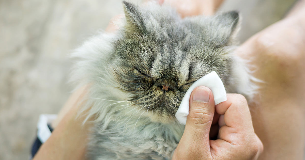
- Wash your hands and use clean, soft gauze or a pet-safe wipe intended for the surrounding area.
- Moisten dried material rather than pulling it from hair.
- Wipe away from the inner corner without rubbing the eyeball.
- Use a fresh surface for each pass and separate material for each eye.
- Pat facial folds dry. Keep long hair from contacting the eye.
- Stop if the pet shows pain or the eye is difficult to open.
Do not use hydrogen peroxide, essential oils, makeup remover, contact-lens disinfectant or a household stain bleach near the eye. Human “get the red out” drops are not a substitute for veterinary assessment.
The 60-second eye record that makes a vet visit more useful
- When it began and whether onset was sudden
- One eye or both
- Color, thickness and amount
- Squinting, rubbing, light avoidance or vision change
- Sneezing, nasal discharge or recent stress in cats
- Recent grooming, shampoo, windy walks, play injury or chemical exposure
- All products already placed in or around the eye
Take a clear photo in the same lighting before cleaning. This preserves evidence that may disappear by the appointment.
Why facial anatomy changes the baseline
Flat-faced dogs and cats, animals with prominent eyes, deep facial folds or hair growing toward the cornea may tear more or retain moisture against the skin. That makes staining common, but it does not make every change “normal for the breed.” Chronic exposure can inflame facial skin, and shallow eye sockets may increase injury risk. A useful baseline is the animal's own stable pattern: sudden increase, one-sided change, pain or a new odor deserves attention.
What veterinarians test when color is not enough
- Fluorescein stain: checks for corneal surface defects and may help assess tear drainage.
- Schirmer tear test: measures tear production when dry eye is suspected.
- Tonometry: measures pressure when glaucoma or inflammation inside the eye is possible.
- Eyelid and lash examination: looks for hairs or anatomy rubbing the cornea.
- Cytology/culture: may be useful in severe, unusual or non-responsive discharge.
These tests explain why two pets with identical green discharge may leave the clinic with different plans. One may have primary conjunctivitis; another may have dry eye with secondary infection; a third may have an ulcer that demands closer monitoring.
Popular tear-stain claims that deserve skepticism
“It is caused by iron in drinking water.”
Tear pigments and chronic wetness are more relevant explanations. Changing water is unlikely to correct a blocked duct, eyelid problem or excessive tearing.
“Antibiotics permanently cure stains.”
Reducing bacteria does not change facial anatomy or remove stained hair. Routine antibiotic use for appearance also raises stewardship concerns.
“Any red-brown stain is harmless.”
Color alone may be cosmetic, but wet, inflamed or odorous skin is not. The eye and the skin beneath it should be assessed separately.
“Wipes treat conjunctivitis.”
External wipes remove debris from fur; they do not deliver treatment to the conjunctiva or cornea.
When eye discharge becomes urgent
Do not wait for a routine appointment if the eye is held closed, cloudy/blue, bulging, bleeding, visibly injured or suddenly unable to see; if pupils are unequal; if a chemical entered the eye; or if pain is rapidly increasing. Prompt examination also matters for yellow/green discharge with redness or discomfort, persistent thick mucus, or a one-sided problem that does not settle.
Choose care by task, not by the word “eye”
The verified Cassiel Pet Eye & Ear Care collection includes formats for external hygiene and more targeted care. Daily wipes belong on surrounding fur and folds. An ophthalmic antibiotic belongs in a separate, diagnosis-dependent path.
If gentamicin has been recommended, current formulation, directions, price and purchase information are on the Cassiel Pet Gentamicin Eye Drops page →
Frequently asked questions
Is brown eye discharge an infection?
Not necessarily. Brown staining often reflects tear pigments and chronic moisture. New odor, redness, pain or thick discharge changes the concern.
Why does my cat have discharge from only one eye?
Local irritation, injury, drainage problems and feline herpesvirus are among many possibilities. Persistent one-sided discharge should be examined.
Can allergies cause green discharge?
Allergy may begin with clear watering, but secondary inflammation or infection can change the discharge. Green color warrants veterinary review.
Can I clean the eye with salt water?
Use a sterile ophthalmic saline product if recommended. Homemade mixtures can have incorrect concentration or contamination and should not be placed in the eye.
Sources
- PetMD: Five Types of Dog Eye Discharge
- Angell Animal Medical Center: Conjunctivitis in Dogs and Cats
- Merck Veterinary Manual: The Conjunctiva in Animals
Educational content, not a diagnosis. Reviewed August 18, 2026.
0 comments